1 LVOT diameter from the PLAX view in most people this is near 2 cm. In general it has excellent correlation with the Gorlin equation.
 To Obtain The Mean Gradient Trace Is Used To Trace The Envelope Of Download Scientific Diagram
The normal aortic valve area is 3-4 cm 2.
Aortic valve gradient calculation. The pressure gradient equals. If a poor or low velocity doppler wave form is displayed reposition the view and move. 1X 2X 3X 4X Aortic Valve Gradient.
Aortic valve area calculation by the Gorlin formula is an indirect method of determining AVA based on the flow through the valve during ventricular systole divided by the systolic pressure gradient across the valve times a constant 443. Management of paradoxical low-flow low-gradient aortic stenosis. This is assumed to be constant throughout systole.
A doppler profile of the aortic valve should be displayed with high velocities in aortic stenosis. 5 Wiggers 6 noted nearly a century ago that significant obstruction to flow occurred when a tube became limited to one third its normal area and this principle is still in use today. The lower the area the higher the gradient P Q2K x EOA2.
Valve area is 10-15 cm2 with a gradient of 25-40 mmHg. The normal aortic valve area is 3-4 cm 2. Delta Pm 8 V2mVpVp Vm where Vp is the peak systolic velocity and Vm the mean systolic velocity.
Valve area is calculated in both the noninvasive and invasive laboratories with the same flow equation. FAV where F is flow A is area and V is velocity so AFV. Regarding Aortic Prosthetic Valves A.
Doppler interrogation of a valve. In low-flow states CO 25 Lmin as the Gorlin equation tends to overestimate the degree of stenosis. In the cardiovascular catheterization laboratory we rely on specific hemodynamic measurements to calculate the size of the aortic valve.
Symptoms tend to not be apparent until the AS is severe. It is more challenging to quantify para-valvular. Valve area is between 15-20 cm2 with a pressure gradient of less than 25 mmHg.
In 20-30 of patients these parameters are discordant usually AVA. The new formula is. The gradient of a mechanical aortic valve is usually around 8 to 22 mmHg which is near the gradient of a normally functioning albeit mildly stenotic natural valve.
Aortic valve area calculation is an indirect method of determining the area of the aortic valve. There are many ways to calculate the valve area of aortic stenosis. Aortic Valve Area LVOT diameter 2 078540 LVOT VTI Aortic Valve VTI CSA LVOT cm 2 0785 x LVOT Diameter SV 0785 x Diameter 2 x VTI LVOT Where LVOT Left Verticular Outflow Tract VTI Velocity Time Integral CSA Cross Sectional Area SV Stroke Volume.
In mild-moderate stenosis the peak-to-peak gradient does not reflect mean gradient however it is often close to mean gradient for severe stenosis. Need for an integrated approach including assessment of symptoms hypertension and stenosis severity. In mild-moderate stenosis the peak-to-peak gradient does not reflect mean gradient however it is often close to mean gradient for severe stenosis.
The below equation relies on the ratio of peak-to-peak instantaneous gradients. Aortic Annulus Size 18-23 cm Mitral Annulus Size 30-35 cm Aortic VTI 18-25 cm. An elevated gradient with a decreased EOA is always suggestive of valvular stenosis C.
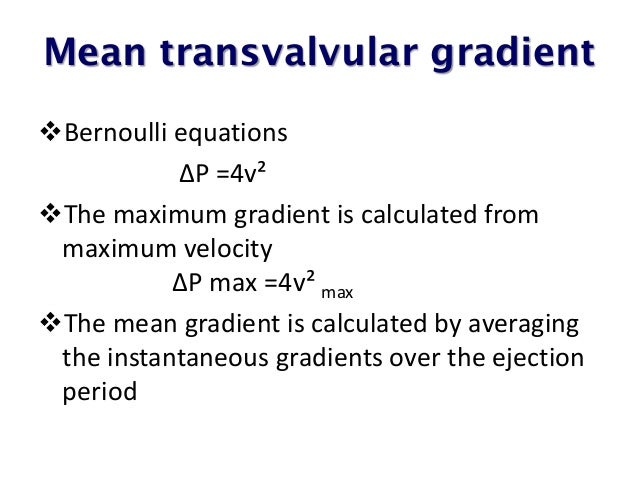
2 LVOT velocity andor VTI from the 5 chamber or apical long axis view. DP is the pressure gradient mmHg across a valve. Assessment of valvular stenosis relies on measurement of the valve gradient and on calculation of valve area.
A gradient of 0 mmHg is normal and usually associated with a normal sized aortic valve. To calculate Aortic Valve Area by the continuity equation you need the following. A routine echocardiogram is required very two years after AVR B.
The calculated aortic valve orifice area is currently one of the measures for evaluating the severity of aortic stenosis. Increase in pressure in the aorta distal to the valve and the vena contracta Decrease in pressure gradient between LV and aorta Increase in EOA distal to the valve and vena contracta Catheter measures distal to the vena contracta Area. Aortic valve area calculation in aortic stenosis by CT and Doppler echocardiography.
This page shows the calculation for aortic valve gradient AVG. In this study the velocity curves in aortic stenosis were analysed mathematically to develop a new and simple method for calculating the mean pressure gradient delta Pm from Doppler velocity tracings. 3 The velocity of VTI at the aortic valve from the 5 chamber or apical.
Transthoracic echocardiogram alone is always sufficient to diagnose valvular stenosis D. Valve area is less than 10 cm2 and with a pressure gradient of greater than 40 mmHg. A valve area of less than 10 cm2 is considered to be severe aortic stenosis.
A critical hemodynamic measurement is the pressure gradient between the left ventricle and the ascending aorta during systole. The most commonly used methods involve measurements taken during echocardiography. A maximum velocity of 4 ms is measured across the aortic valve.
4 4 2 64 mmHg The pressure gradient between the left ventricle and the aorta is 64 mmHg. Echocardiographic assessment of the severity of aortic valve stenosis AS usually relies on peak velocity mean pressure gradient MPG and aortic valve area AVA which should ideally be concordant. For interpretation of these values the area is gene.
Most people with mitral valve prolapse particularly people without symptoms dont require treatment. Mitral valve prolapse is a common cause of a heart murmur caused by a leaky heart valve.
Mitral valve prolapse MVP is a valvular heart disease characterized by the displacement of an abnormally thickened mitral valve leaflet into the left atrium during systole.

What is mitral valve prolapse syndrome. The condition is slightly more prevalent in women than in men. When the mitral valve prolapse condition accompanies symptoms particularly symptoms of dysautonomia it is sometimes referred to as the mitral valve prolapse syndrome or MVPS for short. Mitral valve prolapse is the most common abnormality of the heart valves.
Mitral Valve Prolapse Overview Mitral valve prolapse MVP is a heart condition that affects approximately two percent of the population. The mitral valve which is composed of two flaps allows blood to be pumped from left atrium into the left ventricle of the heart but not back the other way. Most cases of mitral valve prolapse are not serious and only need to be monitored.
The mitral valve is one of the four heart valves. It is the primary form of myxomatous degeneration of the valve. Mitral valve prolapse syndrome MVP is a common condition in which one or both of the flaps cusps of the mitral valve bulge or collapse backward prolapse into the left atrium during ventricular contraction systole.
In a mitral valve prolapse the two. Much of the time MVP doesnt cause any problems. Mitral MY-trul valve prolapse sometimes leads to blood leaking backward into the left atrium a condition called mitral valve regurgitation.
In mitral valve prolapse MVP also. Mitral valve prolapse Angle syndrome Barlow syndrome mesosystolic click and late systolic noise syndrome clapping valve syndrome - deflection protrusion of the valve cusps into the cavity of the left atrium during systole of the left ventricle. Most people with mitral valve prolapse do not have symptoms or signs.
The flaps of the valve are floppy and may not close tightly. There are various types of MVP broadly classified as classic and nonclassic. According to Columbia University Surgery it is nearly 100 that a leaking mitral valve can be successfully repaired.
These flaps normally help seal or open the valve. It is estimated that about 40 of patients with a prolapsing mitral valve experience such symptoms which stem from an imbalance of the autonomic nervous system. Mitral valve prolapse MVP is a heart valve abnormality.
In mitral valve prolapse the mitral valve does not work well because one or both leaflets of the valve are too large which results in an uneven closer of the valve during each heartbeat. Rarely blood can leak the wrong way through the floppy valve. The beverages normally contain carbonated water flavoring and coloring as well as 7-12 percent sugar.
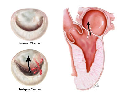
What Are the Symptoms of Mitral Valve Prolapse. Mitral valve prolapse occurs when the flaps leaflets of the hearts mitral valve bulge prolapse like a parachute into the hearts left upper chamber left atrium as the heart contracts. Mitral valve prolapse also known as click-murmur syndrome Barlows syndrome balloon mitral valve or floppy valve syndrome is the bulging of one or both of the mitral valve flaps leaflets into the left atrium during the contraction of the heart.
In some cases this may allow leakage or the backward flow of blood from the left ventricle back into the left atrium mitral. Mitral valve prolapse MVP is a condition in which the hearts mitral valve doesnt work well. Instead they protrude upward into the left atrium the upper left chamber of the heart.
Mitral valve prolapse is a condition in which the leaflets of the mitral valve of the heart do not close normally. When the heart contracts the mitral valve located between the left atrium and ventricle opens to allow the passage of blood into the left ventricleIn mitral valve prolapse the valve has malformations of the leaflets that open and the strings or chordae that support the valveA small amount of blood leaks backward called regurgitation as a result of the leaflets not meeting perfectly. Mitral valve prolapse also known as click murmur syndrome and Barlows syndrome is the most common heart valve abnormality.
If you have mitral valve regurgitation but dont have symptoms your doctor may suggest you return regularly for follow-up examinations to monitor your condition depending on the severity of your condition. Fizzy soft drinks are bad for everyone but theyre especially harmful for men and women with the mitral prolapse syndrome and heart valve disease. Though this may seem like a low percentage the truth is that millions of people live with the symptoms of mitral valve prolapsesometimes without even knowing it.
Your mitral valve which is located between the two is designed to allow blood flow from the left atrium into the left ventricle but not back the other way. That said mitral valve repair is often referred to as the gold standard for treating mitral valve prolapse and Barlows Mitral Valve Disease.